Are you curious about what your eye examination results really mean? Understanding the nuances of your vision health can be a bit overwhelming, but it doesn't have to be! In this article, we'll break down the key components of your eye exam results, clarifying any confusing jargon and helping you grasp the importance of each aspect. So, grab a cup of coffee and join me as we dive deeper into your eye health!

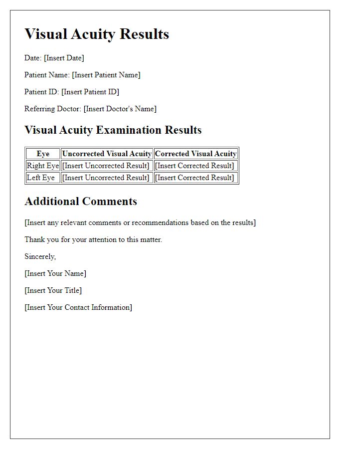
Image cover: Letter Template For Eye Examination Results
Letter Template For Eye Examination Results Samples

Patient Information
Patient information for an eye examination includes critical details such as the individual's name, date of birth, and contact information. The examination may take place at a medical facility specializing in ophthalmology, such as Vision Center or Eye Hospital, where advanced equipment like a phoropter (for measuring refractive error) and an autorefractor (for assessing vision) is utilized. Critical outcomes include visual acuity measurements, often expressed in the Snellen fraction format (e.g., 20/20), and assessments for common ocular conditions like glaucoma or cataracts. A dilated eye exam may be performed, where a mydriatic agent (pupil-dilating eye drop) is applied to enhance visibility of the retina and optic nerve. Follow-up recommendations, including potential prescription for corrective lenses or referrals to specialists, rely heavily on the evaluation of these results.
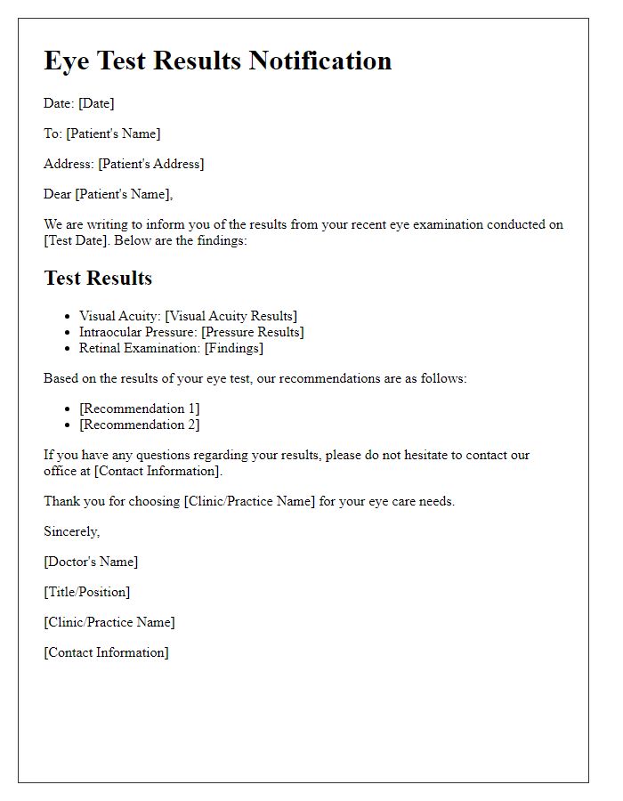
Examination Details
The comprehensive eye examination results reveal essential insights into ocular health and visual acuity. Key metrics include visual acuity readings, measured in Snellen fractions (e.g., 20/20 for normal vision), which indicate clarity of vision at specific distances. The intraocular pressure (IOP) measurement, typically ranging from 10 to 21 mmHg, assesses the risk of glaucoma, while retinal imaging captures detailed visuals of the retina for conditions such as diabetic retinopathy or macular degeneration. Monocular and binocular vision testing highlight differences in each eye's performance and overall coordination, while refractive error determination provides critical information for lens prescriptions (e.g., hyperopia, myopia, astigmatism). The examination incorporates dilated fundus evaluation to review optic nerve health and retinal blood vessels, crucial for diagnosing systemic conditions like hypertension or diabetes. Overall findings guide personalized recommendations for corrective lenses or further treatment modalities.
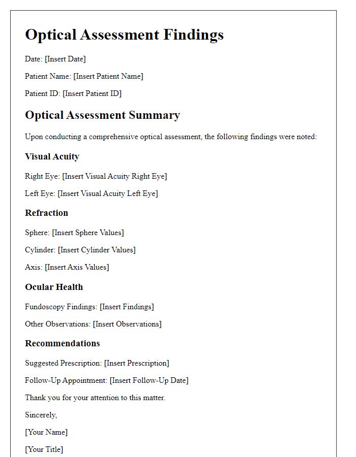
Findings Summary
During the comprehensive eye examination, several key findings were documented regarding visual acuity, ocular health, and potential refractive errors. The visual acuity test revealed measurements of 20/25 in the right eye and 20/30 in the left eye, indicating slightly diminished clarity in the left eye. Additionally, a thorough examination of the retina unveiled early signs of age-related macular degeneration (AMD), particularly in the wet form, which can lead to significant vision loss if not monitored closely. The intraocular pressure was recorded at 18 mmHg in both eyes, falling within the normal range; however, it remains crucial to observe for any changes in future assessments. Furthermore, the evaluation highlighted some astigmatism, suggesting a potential need for corrective lenses for optimal visual performance. Continuous eye care and regular follow-up appointments are essential to managing these findings effectively.
Recommendations
Comprehensive eye examinations reveal critical insights into ocular health. The primary recommendation for individuals identified with conditions like myopia (nearsightedness) or hyperopia (farsightedness) includes regular vision correction, such as prescription glasses or contact lenses, tailored to specific visual acuity needs measured in diopters (e.g., -2.50 for myopia). For those diagnosed with conditions like astigmatism, specialized toric lenses may be necessary to ensure optimal sight clarity. Routine follow-up examinations, typically every 12 to 24 months, are essential for monitoring changes in vision and preventing complications. Lifestyle modifications, including reducing screen time to under two hours daily, implementing the 20-20-20 rule (looking at something 20 feet away for 20 seconds every 20 minutes), and maintaining proper lighting conditions, can significantly alleviate digital eye strain. Additionally, recommending dietary adjustments, such as incorporating dark leafy greens and foods rich in omega-3 fatty acids, supports overall eye health.
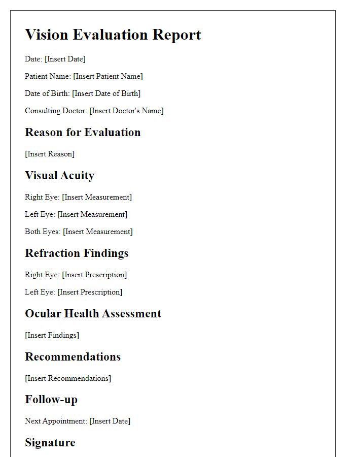
Follow-up Instructions
Following an eye examination, numerous critical factors influence patient care and ongoing visual health. Recommended follow-up appointments are often scheduled within six months to assess any changes in vision quality or eye condition. Patients diagnosed with refractive errors (e.g., myopia or hyperopia) may require updated eyeglass prescriptions, specific to their personal vision needs. Additionally, patients experiencing symptoms such as blurred vision or slight discomfort should monitor their symptoms closely. When necessary, patients may be advised to seek immediate attention in cases of sudden vision loss, indicating potential conditions such as retinal detachment or acute glaucoma, which require urgent intervention. Regular screenings, particularly for individuals over 50 years old or those with a family history of eye diseases, are essential for early detection and prevention of serious conditions.
Read Also: Our Doctor's office's Blogs


Comments